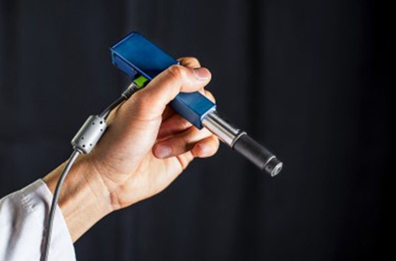
Researchers from the University of Washington have developed a new handheld, pen-sized microscope which could help surgeons identify cancer cells in the operating room and will let them see if tumor cells have been successfully removed or not.
While removing the cancer cells, surgeons have to be very careful about not leaving any cancerous material behind, considering minimum possible neurological harm. Once the operation starts, surgeons don’t have much time. They can’t afford to send tissue samples to a pathology lab, which would take a long time in examining it.
Senior author Jonathan Liu, assistant professor at the University of Washington, said, “Surgeons don’t have a very good way of knowing when they’re done cutting out a tumor. They are using their sense of sight, their sense of touch, pre-operative images of the brain and oftentimes it is pretty subjective.”
But an innovative invention from the University of Washington will revolutionize the way they do these types of operations. This amazing device will allow surgeons to observe their patient on a cellular level, then and there itself.
Liu said, “Being able to zoom and see at the cellular level during the surgery would really help them to accurately differentiate between tumor and normal tissues and improve patient outcomes.”
The Technology Behind This
In this innovative device, “dual-axis confocal microscopy” is used to illuminate and more clearly see through opaque tissue. This device is capable of capturing the details up to a half a millimeter beneath the tissue surface, where some types of cancerous cells originate. It also uses a technique called line-scanning, which scans tissue line by line and also speeds up the image-collection process.
This amazing microscope could be used in a variety of scenarios. Researchers hope that after testing the microscope’s performance as a cancer-screening tool, it can also be introduced into surgeries or clinical procedures.


