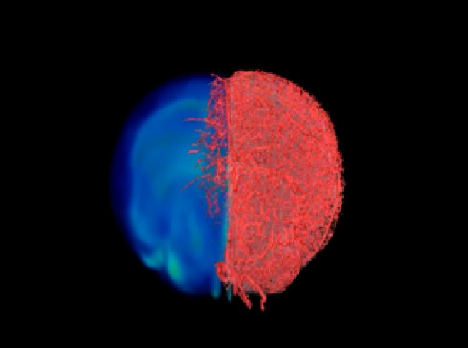
Researchers have developed a new imaging approach that will accelerate imaging-based research in the lab. This approach can help in capturing blood vessel images at different spatial scales.
This 3D visualization method is called VascuViz. Researchers recently tested this technique in mouse tissues. They used a quick-setting polymer mixture that fills up blood vessels and makes them visible in multiple imaging technologies.
Lab tests signify that this technique works well from the largest arteries to the smallest capillaries. The combined images of the blood vessels should also clarify the biology of diseases like cancer and stroke.
It could also improve our understanding of the structures and functions of tissues throughout the body
Mechanical engineer Akanksha Bhargava, from the Johns Hopkins University School of Medicine in Maryland, said, “Now, rather than using an approximation, we can more precisely estimate features like blood flow in actual blood vessels and combine it with complementary information, such as cell density,”
This imaging approach can help answer important questions in the broader field of micro-circulation
VascuViz-based measurements can be entered into computer simulations of blood flow to discover the way blood is flowing. As a result, this information can help better understand how diseases work and might be progressing.


