Since the beginning of medicine, the best way of learning about human anatomy, especially the brain, was using fresh cadavers to dissect. Throughout the 20th century, new brain scanning technologies have been developed and allow us to look at the structure and function of the brain while it’s still working. However, we are still learning a lot from the deceased brain with scanning technologies.
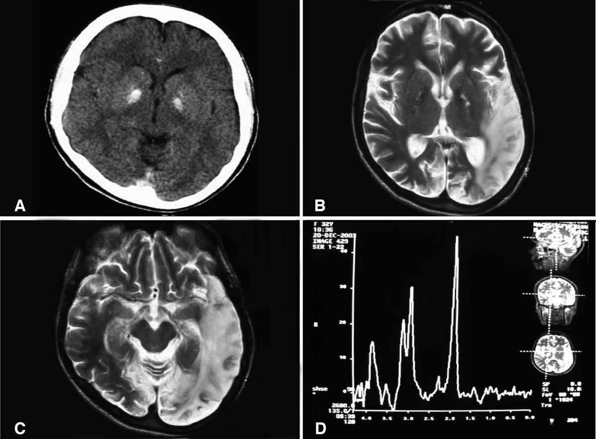
Forensic pathologists are keen on using post-mortem brain scanning to determine the cause of death and understand the lethal effects of brain damage. A blunt blow to the head, internal bleeding, stroke, or a brain aneurism are difficult to identify without this technology. Autopsies are time consuming and often impractical for the victim’s family who would like to have an open casket funeral.
A growing number of studies are also looking at more subtle effects by comparing the size and shape of brain structures at death in people who were diagnosed with conditions such as schizophrenia or Alzheimer’s disease. It is still possible to use traditional techniques for completing these studies, such as a camera and a scalpel, or even more advanced approaches such as mechanically cutting the brain into micro-thin slices and digitally photographing each section. But using a standard brain scanner is quick, cheap and gives scientists the option of using the same software developed for analyzing living scans.
But some of the most interesting neuroscience studies on the dead are not aimed at understanding mortality, but are focused on tuning technologies for the living. One of the most popular brain imaging technologies, called functional magnetic resonance imaging or fMRI, involves detecting tiny differences in radio signals emitted from brain tissues and blood as their hydrogen molecules are manipulated by large magnetic fields.
The only problem reading the data from fMRIs is that they can be easily interfered with by the other functions of the body like breathing or body movement, and it’s hard to distinguish between the occurrences. But as dead people do little moving and breathing, they make perfect specimens for the scans making it easy to show the flaws in the scanning technology. For instance, if the scans had shown a part of the brain that was still active in a deceased person and picked up anomalies that were repetitive in comparison. Using the deceased as a control helps point out the flaws for when the scans apply to the living.

